

When assessing necrotic tissue and eschar, granulation tissue, or slough in wound care, start with what you're actually seeing. A wound can contain more than one tissue type, and some healthy structures can be mistaken for slough or nonviable tissue. Document the tissue present, what was removed during debridement, and the depth reached. The record must support both the clinical assessment and the code submitted.
Misidentifying tissue in a wound bed isn't a minor documentation issue. It changes what you do next.
A healthy structure can be mistaken for slough and inadvertently removed. When stable eschar is treated without sufficient attention to the underlying clinical picture, it can lead to problems. And when the tissue described in the note doesn't match the tissue actually removed, the documentation may not support the procedure code submitted.
In addition to prolonged stress on the patient, you might also struggle to get your claim approved.
This is why, as you know, correct tissue identification is imperative, especially when a wound is being prepared for a skin substitute graft. While our application technique checklist covers the application process, you first need a clear picture of the wound bed you're working with.
Granulation tissue and slough are two common findings, but they're not the only tissue types you'll encounter. Necrotic tissue and eschar also need to be distinguished from viable tissue, and a single wound can contain more than one tissue type.
Here's what to look for.
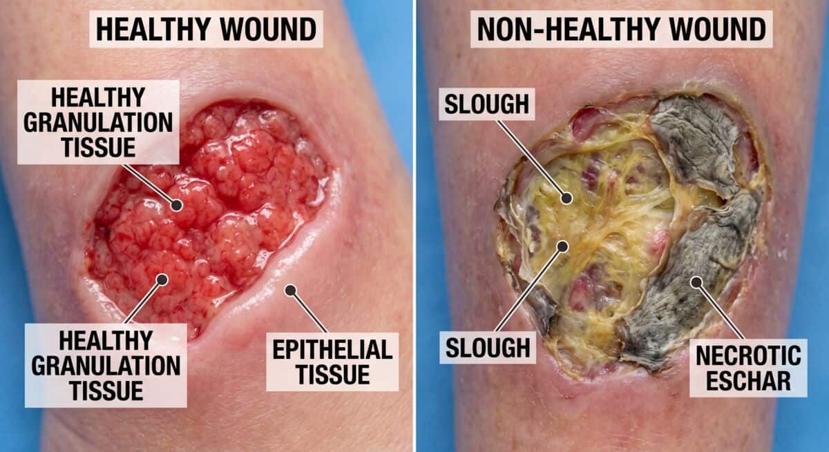
Necrotic tissue is nonviable tissue. It can result from factors such as inadequate blood supply, infection, or trauma.
Eschar is one possible presentation. It may appear dry, dark, and firmly attached to the wound bed. However, not every area of eschar should automatically be debrided. The decision depends on the wound, the patient's condition, and the clinical findings.
Stable, dry eschar without signs of infection may be managed differently from eschar that is separating, draining, or associated with other concerning findings. If vascular status is a concern, that needs to be assessed as part of the clinical decision-making process.
A simple visual label isn't enough. Document what you see and the factors that support the treatment decision. If debridement is performed, document the tissue actually removed and the depth reached.
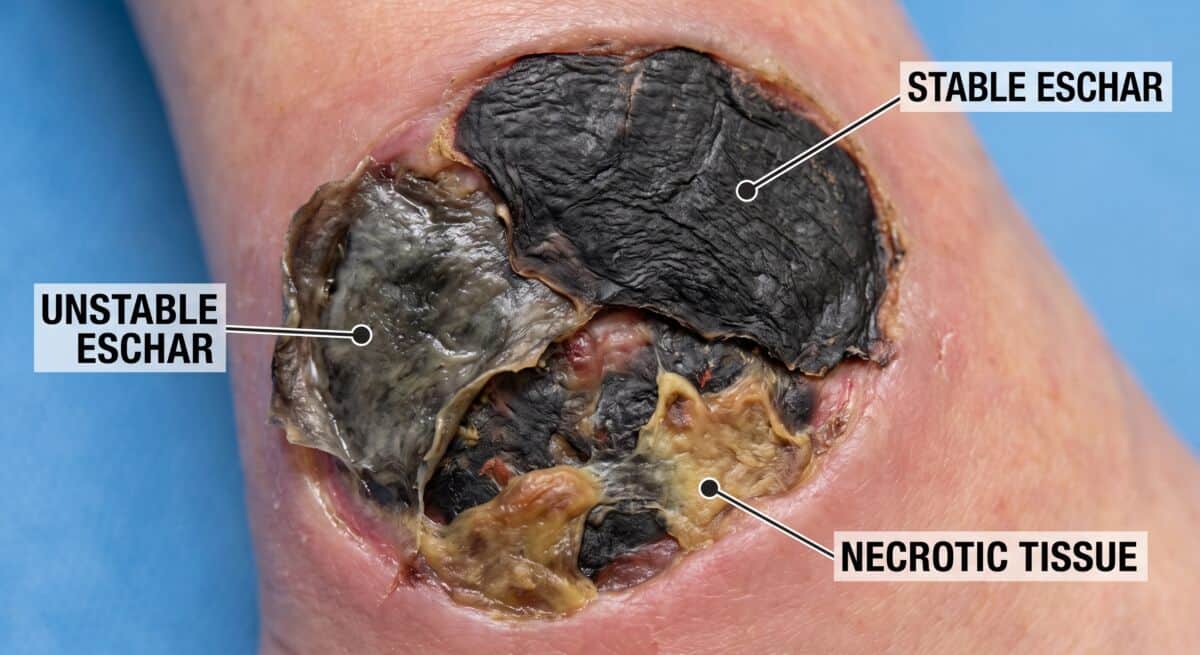
Slough can appear yellow, tan, gray, or green and may be loose, stringy, or firmly attached to the wound bed. However, it can be difficult to distinguish from other pale or fibrous structures in some wounds.
If you're unsure of what you're looking at, don't let the label drive the procedure. The clinical assessment comes first.
The appropriate approach to slough also depends on how it presents and the patient's overall condition. Treatment may involve different forms of debridement or other wound-bed management strategies.
Chart documentation is rather straightforward:
If you're tracking the wound over multiple visits, documenting the tissue composition consistently makes changes in the wound bed easier to see.
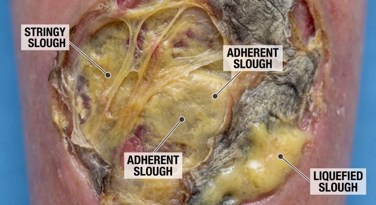
This is one of the more important distinctions to make at the bedside. Some healthy structures can look pale, fibrous, or otherwise similar to slough. Tendon, fascia, muscle, fat, and other tissue can be misidentified when the wound is difficult to assess.
The open-access Tissue Types chapter from Skin and Wound Care for Health Care Professionals provides additional guidance on distinguishing these structures. If you're unsure what you're seeing, don't let a label in the assessment determine what’s removed.
The clinical assessment always comes first.
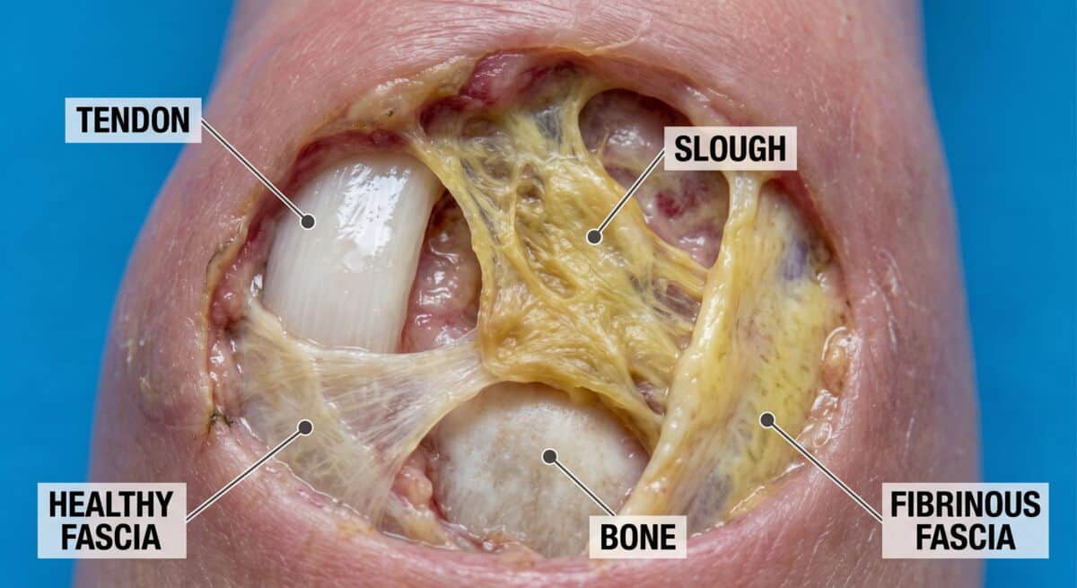
CMS guidance ties debridement coding to the tissue removed rather than the deepest part of the wound. Selective debridement codes 97597 and 97598 and surgical debridement codes 11042–11047 apply to different services and tissue levels.
CPT 97597 and 97598 shouldn't be selected simply because the wound contains slough.
Your documentation must support the code reported. The code must always match the service actually performed.
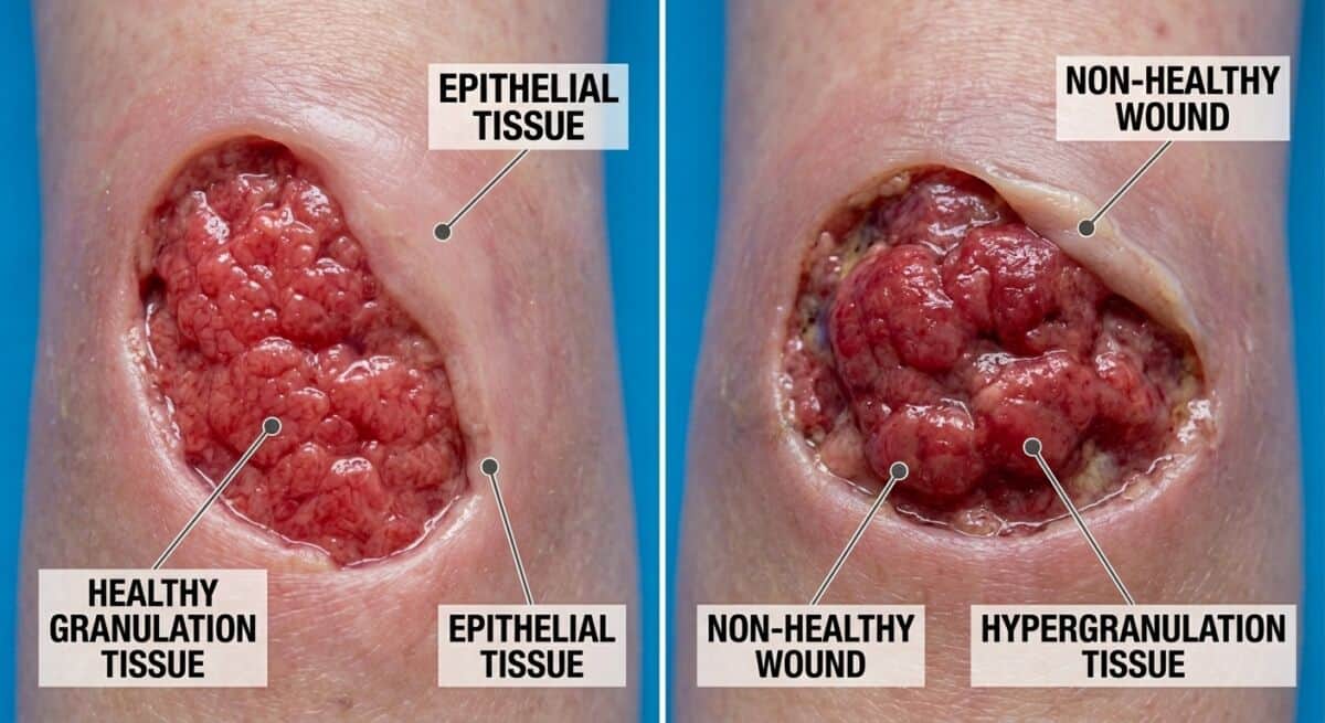
Healthy granulation tissue is generally pink to red, moist, and granular in appearance. It's a normal part of wound healing and can become increasingly prominent as a wound progresses.
When documenting it, describe what you actually see rather than simply writing "healthy wound bed." Also explain how it compares with previous assessments.
For example:
The amount and appearance of granulation can change as the wound progresses, so documenting it consistently makes comparisons between visits more useful.
CMS wound-care guidance notes that surgical debridement isn't considered reasonable and necessary when the record describes pink-to-red granulation tissue without devitalized, fibrotic, or nonviable tissue. That doesn't mean the presence of granulation tissue automatically rules out every type of debridement.
Your documentation should describe the tissue present and support the procedure that was performed.
Hypergranulation isn’t healthy granulation. It can extend above the level of the surrounding wound edge and may be friable or prone to bleeding. When it appears, the underlying cause and appropriate treatment need to be assessed clinically.
The important point for documentation is to distinguish healthy granulation from tissue that is abnormal or interfering with wound progression.
|
Tissue type |
Typical appearance |
What to document |
Treatment / coding consideration |
|
Necrotic tissue / eschar |
Can be dark, dry, leathery, or otherwise nonviable |
Location, appearance, wound characteristics, and relevant clinical findings |
Treatment depends on the wound and patient. If debridement is performed, document the tissue actually removed and depth reached. |
|
Slough |
Often yellow, tan, gray, or green; may be loose or adherent |
Tissue type, extent within the wound bed, and relevant wound characteristics |
If selective debridement is performed, documentation should support the service and code selected. |
|
Granulation tissue |
Typically pink to red, moist, and granular |
Presence, extent, and changes over time |
Document the wound's current state and whether other nonviable tissue remains. |
|
Hypergranulation |
Raised above the surrounding wound edge; may be friable |
Appearance, extent, and relevant clinical findings |
Clinical management depends on the underlying cause and individual wound |
The table is a reference, not a substitute for clinical assessment. Wound appearance can vary, and more than one tissue type may be present at the same time.
The depth of the wound isn't necessarily the depth of the debridement. Your record should identify the tissue that was actually removed and the depth reached.
CMS's current wound-care guidance states that surgical debridement codes should reflect the level of tissue removed, not the deepest structure visible in the wound.
For example, exposed bone doesn't automatically support a bone-debridement code if bone wasn't actually removed. Your chart must make that distinction clear. Similarly, if muscle or fascia is debrided, the record needs to support that level of treatment.
CMS's wound-care guidance also states that selective debridement codes 97597–97598 shouldn't be reported with surgical debridement codes 11042–11047 for the same wound on the same date of service.
Always check if the procedure note clearly support the code that was submitted. If it isn't obvious, the documentation may need closer review.
Your documentation doesn't need to become a long narrative. However, a useful wound assessment gives the next person reviewing the chart enough information to understand what was present and what changed.
Depending on the service and applicable payer requirements, that may include:
CMS's current wound-care guidance calls for documentation of wound characteristics, including dimensions, depth, tissue type, and other relevant findings. It also expects the record to support the type and depth of debridement performed.
Chronic wound photos certainly help support your care records, but they don't replace a useful written assessment.
If a skin substitute graft is applied, the record should support the wound's condition and the application performed. CMS guidance for applicable skin substitute applications calls for wound characteristics to be documented throughout the episode of care.
Before a skin substitute graft is applied, your team needs a clear picture of the wound's current condition. The record should show what’s been treated, what remains, how the wound has changed, and that it’s healthy enough for skin graft treatment.
According to CMS guidelines for appropriate skin substitute applications, you must record the wound's characteristics before, after, and during all stages of the wound care.
Consistent, comprehensive wound assessments don’t just improve your chances of claim approval; they also help with clinical decision-making.
RenewMed's role is to support providers on the administrative side of Medicare’s wound care processes.
Through our White Glove Service, we, among other support,
We’re here to help lessen your load and ease your wound care decisions.
Let’s Discuss Medicare’s Wound Care Specifics
No. A photo adds useful visual information, but it doesn't replace the written record.
Your documentation still needs to describe the wound and the procedure performed. Photos are most useful when they're consistent enough to help you compare the wound over time.
Yes. Accurate wound-bed assessment helps document the condition of the wound and its progression. For applicable skin substitute applications, CMS guidance calls for wound characteristics to be documented throughout the care episode.
Granulation is one part of wound-bed assessment. It shouldn't be treated as the sole measure of whether a wound is appropriate for a skin substitute. You should assess the wound and the patient as a whole and make the treatment decision based on the patient's clinical circumstances.
Don't force a tissue label simply to complete the documentation. Follow your clinical protocols and obtain appropriate clinical input when the tissue cannot be confidently identified.
A wound can change between visits, and more than one tissue type can be present at once.
A useful record is the one that describes what the wound looked like, what was treated, how and why that treatment was chosen, and how the wound has changed. That makes the clinical record easier to follow and gives the billing team a clearer basis for the code submitted.
Good documentation keeps the pieces connected, one patient at a time.
Contact Your RenewMed Consultant
Disclaimer: This content is created for licensed healthcare professionals, offering educational insights into wound care. It is not intended as medical advice or to replace your own clinical judgment when treating patients. We're here to support you, but the final treatment decisions should always be based on your professional evaluation of each unique patient's needs.
