The staging system for pressure injuries (bedsores) looks simple on paper. You have six categories. You learn clear definitions. You memorize visual criteria.
But bedside reality is much messier.
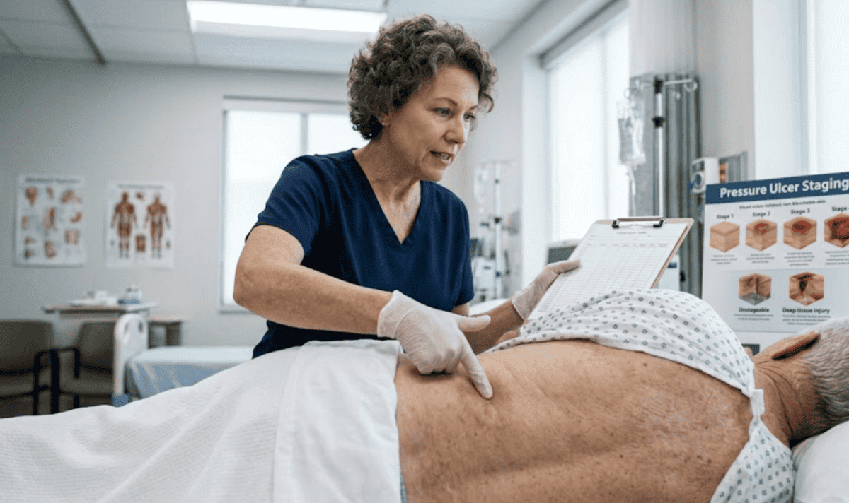
You're looking at damage that doesn't fit neatly into a textbook illustration. The patient's history is complicated. Yet, your documentation MUST be precise for clinical and reimbursement purposes.
Your treatment path forward depends on getting your assessment right. So, this pressure ulcer staging guide is for providers who treat Medicare patients with chronic wounds.
Accurate staging drives everything that follows.
Get it wrong, and you're potentially delaying healing for a patient.
RenewMed focuses on removing the administrative friction that often follows a complex diagnosis. We handle the hours spent on hold for prior authorizations and the complexities of IVRs, so you can focus on the clinical nuances of tissue repair.
Evaluating chronic wounds is rarely straightforward. This pressure ulcer staging guide outlines the clinical markers for Stage 1 through Stage 4, exploring the complexities of unstageable wounds and the risks of documentation errors. Its goal is to help you make confident, compliant treatment decisions.
Accurate staging requires an understanding of what is happening beneath the skin's surface.
This guide serves as a framework for your daily rounds, helping you distinguish between surface-level friction and true structural damage.
Source: Sturdy
Stage 1 pressure injuries are the easiest to miss. They're also the most important to catch.
The definition seems simple: Intact skin with non-blanchable erythema. Press on the area, release, and the redness doesn't fade.
In patients with lighter skin tones, this presents as a distinct red patch. In patients with darker skin tones, redness may not be visible at all. This is where many providers struggle.
Source: The Clinical Excellence Commission
In patients with darker skin tones, look for a persistent purple, ashy, or maroon tint, or rely on the "feel" of the tissue.
You’re looking for a change in the skin’s "behavior" compared to the surrounding area. This may present as:
This is vital because Stage 1 is your intervention window. The skin is still intact. You aren't managing an open wound yet. At this stage, you can often prevent progression entirely.
The goal is to offload the area and stop the progression to an open wound.
Key interventions include:
Stage 1 doesn't require aggressive wound care products or debridement. These injuries often resolve with simple pressure relief. Document them even when the patient downplays the severity.
These are early warning signs. They establish a timeline that becomes critical if the wound progresses.
Stage 2 marks the transition to an open wound. You're looking at partial thickness tissue loss involving the epidermis and probably the dermis. The wound bed is typically pink or red and moist. You may see an intact or ruptured blister.
This is where staging errors start to multiply.
Source: The Clinical Excellence Commission
One of the most common mistakes is misdiagnosing Stage 2 pressure ulcers. Unfortunately, it’s easy to confuse a Stage 2 ulcer with other skin conditions.
Stage 2 wounds are generally manageable with conservative care.
Most Stage 2 pressure ulcers should show signs of closure within two weeks.
However, if the wound edges don't show closure movement after 14 days, it may be entering a chronic, stalled state. At this point, review the patient’s systemic health (uncontrolled diabetes or poor peripheral circulation) that might be preventing healing.
If you don't see progress after 30 days of appropriate and documented care, it meets the definition of a chronic wound. Your patient should benefit from an advanced wound care consultation.
In a Stage 3 ulcer, the skin loss is full thickness.
You’ll see adipose (fat) tissue in the wound bed. You may also see slough (yellow/tan stringy tissue), epibole (rolled wound edges), or eschar (dry, black, necrotic tissue), but they don't obscure the wound’s depth. The bone, tendon, and muscle aren't exposed yet.
Clinical assessment becomes much more demanding here.
Pressure Ulcer Stage 3
Source: The Clinical Excellence Commission
Stage 3 wounds are deceptive because of “tunneling” and "undermining." The visible hole on the surface might be small, but the damage can extend far beneath the skin edges.
Use a sterile swab to "map" the edges of the wound. If you find pockets where the swab goes under the skin, you’re dealing with undermining. If the swab finds narrow, deep tracts that formed in one direction, it’s tunneling. These significantly increase the risk of an abscess forming.
Stage 3 wounds also vary significantly in depth based on anatomical location. A Stage 3 wound on the sacrum may be quite deep. A Stage 3 wound on the bridge of the nose may appear relatively shallow. Both represent full thickness loss.
Don't stage based on perceived severity. Stage based on the anatomical structures you observe. If you see fat but not fascia, muscle, or bone, you're likely looking at Stage 3.
This is the stage where standard dressings fail to provide enough biological "stimulus" for healing. You also face a higher risk of infection with full thickness wounds.
You need to apply a more aggressive wound management approach.
Stage 4 is full thickness skin and tissue loss with exposed or directly palpable fascia, muscle, tendon, ligament, cartilage, or bone.
These wounds consume clinical resources and test your treatment plans daily.
Pressure Ulcer Stage 4
Source: The Clinical Excellence Commission
Stage 4 ulcers are a clinical emergency.
The exposed bone puts the patient at an extremely high risk for osteomyelitis (bone infection). You aren't just managing a wound but a systemic threat. Clinical assessment alone is no longer enough. Treatment usually requires a surgical consultation and intensive nutritional support.
Even with optimal care, Stage 4 pressure wounds take months to heal. So, manage patient and family expectations accordingly.
Once any infection is cleared and the wound bed is granular, advanced biologics act as a "bridge" to closure.
Amniotic membrane grafts provide extracellular matrix components that support tissue regeneration. They also help protect exposed structures and reduce the chronic inflammation that characterizes these deep wounds. Most importantly, they significantly reduce the overall healing time.
A wound is "unstageable" when you can't see the base because it's covered by slough or eschar. You know it’s at least a Stage 3 or 4, but you can't confirm the exact depth until the debris is removed.
Unstageable Pressure Ulcer
Source: The Clinical Excellence Commission
It’s tempting to "guess" the stage based on the patient's history, but this is a documentation trap. Until the wound is debrided, it must remain "unstageable" in your records. But add notes about the eschar characteristics of the wound.
Guessing can lead to audit risks and mismanaged treatment expectations.
If the eschar is dry, adherent, and shows no redness or fluctuance, it acts as the body's natural cover.
In most cases, it’s best to leave it alone.
However, for most other sites, debridement is necessary to reveal the true extent of the injury and prepare the site for advanced healing solutions.
Once the necrotic tissue is removed, the wound must be restaged immediately. This is the moment where your treatment plan pivots.
If debridement reveals a clean Stage 3 base, it becomes a prime candidate for amniotic grafting.
The transition from "unstageable" to "prepped for a graft" involves a lot of moving parts. Our White Glove Service is designed for this. Once you’ve prepped the wound, we handle the logistics.
Accurate staging is a skill that improves with every patient, but the administrative burden shouldn't have to. RenewMed exists to bridge the gap between your clinical expertise and the operational demands of modern wound care.
More than products, we provide the business intelligence and back-office support you need to keep your practice profitable while delivering the highest standard of care.
SOFT CTA: If you’re ready to streamline your wound care program, connect with RenewMed today.
Can a pressure ulcer be down-staged as it heals?
No. Once a wound has been staged, it retains that stage throughout healing.
A Stage 3 wound that's healing doesn't become a Stage 2 wound. It's documented as a "healing Stage 3 wound." Regenerated tissue is scar tissue, not original anatomical structure.
Can I stage a wound if I only see slough in part of it?
If the slough covers the base so that the true depth can’t be determined, it is unstageable. If you can clearly see the depth despite some slough, you can stage it.
What if I see exposed bone but the wound is small?
The size of the wound does not dictate the stage; the depth does. If bone is palpable or visible, it is a Stage 4, regardless of the surface diameter.
How do I distinguish deep tissue pressure injury from Stage 3?
Deep tissue pressure injury (DTPI) appears as a persistent, non-blanchable deep red, maroon, or purple discoloration. It represents damage that originated in deeper tissues.
It looks like a bruise but indicates damage to the muscle-bone interface. These often evolve rapidly into Stage 3 or 4 ulcers. So, monitor them very closely.
When should advanced wound care products be used in pressure ulcer staging?
The 30-day threshold is the best guidance. If a wound hasn't reduced in size by 20% to 50% after 30 days of conservative care, it's a chronic wound. At this point, consider skin substitute grafts.
Earlier intervention may be appropriate for wounds with severe prognostic factors.
What documentation is essential for pressure ulcer staging?
You need the wound location, stage, exact measurements, and wound bed description. Note the periwound skin condition and exudate characteristics. Adding in photos of the wounds helps support your case.
For Medicare patients, documenting the wound duration is absolutely critical for reimbursement.
Does RenewMed help with the Q-code and billing process?
Yes. The 2026 Medicare fee schedule shifted many skin substitutes to a flat "incident-to" payment rate of approximately $127.14 per square centimeter.
Part of our White Glove Service includes evaluating your formulary to ensure you have access to clinically effective grafts that remain financially viable under this new financial cap. We also guide you on specific documentation protocols to protect your practice from clawbacks.
Your focus should be on the wound bed, not the insurance portal. Join us in making a difference for your patients by streamlining your access to the world's most advanced healing biologics.
Every healed wound is a life changed. We're here to help you make that difference.
Sources used:
Disclaimer: This content is created for licensed healthcare professionals, offering educational insights into wound care. It is not intended as medical advice or to replace your own clinical judgment when treating patients. We're here to support you, but the final treatment decisions should always be based on your professional evaluation of each unique patient's needs.