
Lower extremity wounds rarely fail because of dressing selection. They fail because the underlying cause wasn’t identified early enough.
You need a decisive lower extremity wound assessment to determine if you're dealing with a drainage problem (venous) or a delivery problem (arterial).
What separates the providers who consistently heal these wounds isn't just access to advanced wound care products but the depth of their initial assessment. When you understand the full picture, you can match the right therapy to the right wound.
A successful treatment plan starts with a precise diagnosis. This lower extremity wound assessment moves beyond surface-level documentation to uncover the vascular and biological drivers of chronic leg and foot ulcers. We focus on differentiating arterial and venous disease and identifying the "tipping point" for advanced interventions.
An effective leg and foot ulcer assessment is a diagnostic framework that ensures you aren't treating a symptom while ignoring the cause.
By systematically evaluating the vascular supply, the wound bed, and the health of the surrounding skin, you can pinpoint the precise "tipping point" where a wound stalls. This structured approach enables you to create a decisive treatment plan, ensuring that every intervention, from compression to advanced biologics, is backed by clinical data.
The most dangerous mistake in wound care is applying compression to a leg with undiagnosed arterial disease. Before you even touch the wound bed, you must look at the limb as a whole.
You’re not merely looking for vascular disease but at how that disease is starving or drowning the tissue.
Arterial wounds starve the tissue of oxygen. These typically appear on the toes, the top of the foot, or the lateral malleolus.
They're often dry, "punched-out," and incredibly painful. If the skin is shiny, hairless, and cool, you're likely looking at an arterial issue.
The hallmark sign? Pain that worsens when the leg is elevated (rest pain).
Venous ulcers are about stagnation. Blood pools, causing edema and "hemosiderin staining" (that brownish discoloration). You'll also see irregular borders and heavy exudate.
These wounds usually appear in the "gaiter" region (ankle to mid-calf).
The patient often reports a dull ache that improves with elevation.
Many Medicare patients have both. If you apply heavy compression to a leg with unrecognized arterial disease, you risk causing further tissue necrosis.
Always verify perfusion before you squeeze.
If these are present in an acute setting, you aren't looking at a chronic wound. You are looking at a vascular emergency.
If the patient presents with Pulselessness and Pallor, the wound bed is secondary; the priority is a STAT vascular referral.
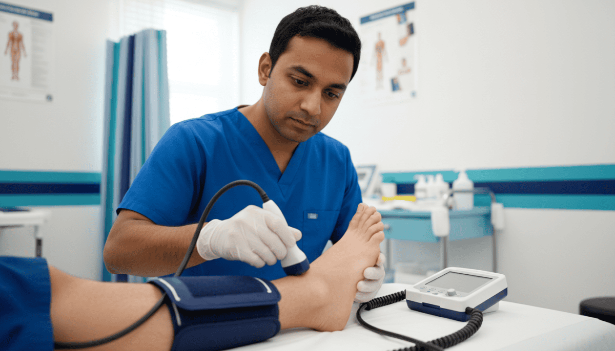
The Ankle-Brachial Index (ABI) is the gold standard for your initial lower extremity wound assessment. It’s a simple ratio of the systolic blood pressure at the ankle compared to the arm, but it dictates your entire safety profile for treatment.
It tells you if the arteries can deliver the blood needed to support the metabolic demands of healing.
|
ABI Score |
Interpretation |
Clinical Action |
|
0.9 – 1.4 |
Normal |
Proceed with standard care and compression (30-40 mmHg) if needed. |
|
0.7 – 0.9 |
Mild Peripheral Artery Disease (PAD) |
Use caution with compression; monitor closely. |
|
0.5 – 0.7 |
Moderate PAD |
Refer to Vascular. Only use light compression. The wound is at high risk for stalling. |
|
< 0.5 |
Severe Ischemia |
DO NOT COMPRESS. High risk for necrosis/amputation. Revascularization needed. Advanced grafts are contraindicated until blood flow is restored |
|
> 1.4 |
Calcified Vessels |
Inaccurate reading (common in diabetes); use Toe-Brachial Index (TBI). |
In patients with diabetes, medial arterial calcification can cause falsely elevated ABIs. A "normal" 1.30 might actually be a severely compromised vessel that's too stiff to compress.
If the clinical picture doesn't match the number, get a TBI.
Once you've cleared the vascular checks, it's time to look at the wound itself. There are a few lower extremity wound assessment models you can use here.
The skin surrounding the wound is a window into your moisture management strategy. During your lower extremity wound assessment, pay close attention to:
If the periwound is failing, the wound won't close. It’s that simple.
Every chronic wound has bacteria, but not every wound is infected. What this means is that although all chronic wounds are "colonized" (bacteria are present but not causing harm), you only treat "infections" (where bacteria are invading tissue).
So, how do you know if you should start the patient on antibiotics? Simple. Look for the "NERDS" and "STONES."
If you see "STONES," you're dealing with a deep-seated issue that requires systemic intervention. If it’s "NERDS," local biofilm management and advanced grafts are often enough to turn the tide.
This is the point where treatment delays usually happen: waiting on approvals, documentation, and product access. RenewMed steps in here to remove that delay so you can act immediately.
Your best assurance to get a claim approved is to make sure your clinical notes are comprehensive from the start. It might feel like a hassle to do this at every visit, but getting into this habit makes a huge difference.
The 7 Basic Parameters of Wound Assessment
Standardized documentation is your best defense against audit risks. Every note should include:
RenewMed Tip: Include clear, dated photos of the ulcer taken at each visit to substantiate and double-check your notes.
We know that the "administrative load" of wound care starts during assessment.
You and your team have questions, like: If your ABI is 0.75, does the payer require a vascular consult before they’ll approve a graft? If the wound is 0.8cm², is it still eligible?
Our White Glove Service is designed to handle these questions for you. We provide the business intelligence and documentation support to ensure your leg and foot ulcer evaluation findings are translated into a compliant, approved treatment plan.
We manage the IVRs, prior authorizations, and product logistics. So when you decide a patient needs an advanced biologic, the product is ready and your revenue protected.
f your lower extremity wound assessment shows a clean, granulating bed that has stalled for 30 days, call your RenewMed consultant.
Follow this 5-minute sequence to ensure no critical data point is missed.
What is the difference between colonization and local infection?
Colonization is the presence of bacteria that aren't causing tissue damage.
Local infection (often signaled by increased exudate or friable tissue) means the bacteria are actively interfering with the healing process and require intervention.
How often should a lower extremity wound assessment be performed?
A full, documented assessment should happen at least weekly. However, the wound should be inspected at every dressing change to check for signs of rapid deterioration or new infection.
Can I perform an ABI if the patient has a painful ulcer?
It can be difficult. If the cuff pressure is too painful, consider a Toe-Brachial Index (TBI) or a bedside arterial Doppler to assess phasicity (triphasic vs. monophasic sounds) as an alternative.
Is edema a contraindication for grafting?
No, but it must be managed. If the leg is "weeping" fluid, the graft will not adhere. Edema must be controlled via compression (if ABI >0.5) or elevation before the biologic is applied.
When does RenewMed's clinical team get involved?
Ideally, as soon as you have your vascular baseline. We can help you match the specific tissue type in your assessment to the most effective graft in our portfolio, ensuring a higher likelihood of first-time closure.
Stop letting the "documentation hurdle" keep you from providing the best care.
Every inch toward closure is a step toward a saved limb. We’re with you in that mission.
Sources used:
Disclaimer: This content is created for licensed healthcare professionals, offering educational insights into wound care. It is not intended as medical advice or to replace your own clinical judgment when treating patients. We're here to support you, but the final treatment decisions should always be based on your professional evaluation of each unique patient's needs.
